Mr. Majestic's Microscopy..... and life on Mars?
Message boards :
Cafe SETI :
Mr. Majestic's Microscopy..... and life on Mars?
Message board moderation
Previous · 1 · 2 · 3 · 4 · 5 . . . 8 · Next
| Author | Message |
|---|---|
 Mr. Majestic Mr. Majestic Send message Joined: 26 Nov 07 Posts: 4752 Credit: 258,845 RAC: 0 
|
Know if only I had the money for this..... Yes, this image is an SEM (scanning electron microscope). I have no idea what the highest magnification on this rig is, it's not the one I found for sale, but I know they can go up to 250,000 times magnification. And to think, mine only goes up to 1250!   ID: 756654 · |
|
Mr. Majestic Send message Joined: 26 Nov 07 Posts: 4752 Credit: 258,845 RAC: 0
|
Here is the bacteria that causes strep throat (strepto-coccus): 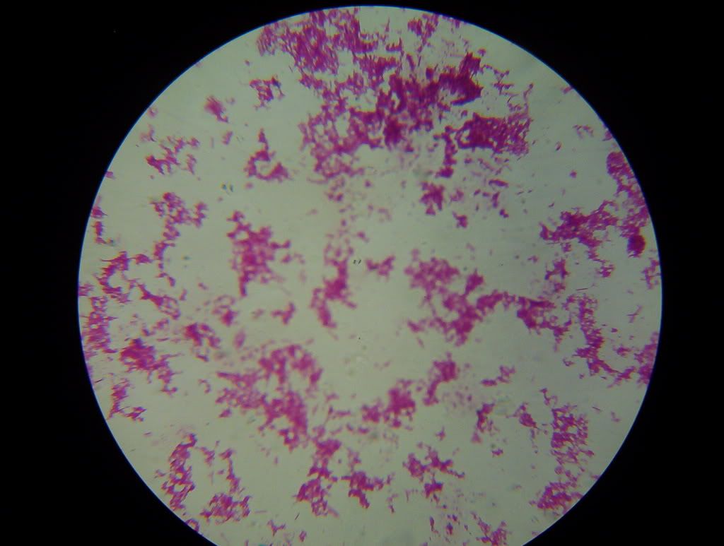 ID: 756657 · |
|
Mr. Majestic Send message Joined: 26 Nov 07 Posts: 4752 Credit: 258,845 RAC: 0
|
Here is the bacteria that causes strep throat (strepto-coccus): Sorry about the low image quality. The image didn't turn out as good as I thought it had. I will try to get a better one later. ID: 756658 · |
|
Beethoven Send message Joined: 19 Jun 06 Posts: 15274 Credit: 8,546 RAC: 0 |
Know if only I had the money for this..... Only huh? Heh. I remember when I was a kid, that we had this marsh swamp not to far away from home. I'd bring back some swamp water and put a drop under a 600x magnification microscope I had. It was just fascinating to watch those paramecia, amoebas and well, just weirdo creatures I never found the names for. It was like watching an aquarium from outer space. Great fun!!! :]] That strep throat bacteroa looks as nasty as it is. LOL ID: 756675 · |
|
Mr. Majestic Send message Joined: 26 Nov 07 Posts: 4752 Credit: 258,845 RAC: 0
|
Know if only I had the money for this..... That is how my obsession started. I got a $30 microscope with plastic objective lenses and decided that I wanted to learn more about what I was seeing. I got some books on microbiology and eventually worked my way up to a $500 scope and some college text books. This and the high number of heart related deaths in my family is part of the reason I want to be an open heart surgeon. It is amazing what you can see with this thing. I will try to get some pictures of E-coli, staph, and others to turn out to post them. I will also try to get a view of the strepto-coccus at 1250x magnigication. ID: 756679 · |
|
Dune_Finkleberry Send message Joined: 25 Feb 06 Posts: 6454 Credit: 198,656 RAC: 0
|
That is how my obsession started. I got a $30 microscope with plastic objective lenses and decided that I wanted to learn more about what I was seeing. I got some books on microbiology and eventually worked my way up to a $500 scope and some college text books. This and the high number of heart related deaths in my family is part of the reason I want to be an open heart surgeon. It is amazing what you can see with this thing. I will try to get some pictures of E-coli, staph, and others to turn out to post them. I will also try to get a view of the strepto-coccus at 1250x magnigication. I hope I never meet you on the business end of a scalpel. Cheers AE! Enjoying your pics. Account frozen... ID: 756734 · |
|
Clyde C. Phillips, III Send message Joined: 2 Aug 00 Posts: 1851 Credit: 5,955,047 RAC: 0
|
I see that those adapters only cost about $50US or so. Do they adapt a camera to the third (photographing) head of a trinocular or to one of the viewing eyepieces of a binocular (or monocular) microscope? The camera that Dr Ceti produced exactly exemplifies what I've seen: Just 470 TV lines for $700. No wonder! It's a video camera! In todays paper I see a new Nikon Coolpix P80 still that has 10.1 (effective) megapixels (one needs to compare apples and oranges here) and an 18x zoom for only $370. Wonder how sharp that lens is. Wonder whether it is fully achromatic or has a purple fringe. Wonder where along the focal lengths that lens performs at its best. Wonder if that camera could be adapted to a binocular microscope without appreciable image degradation. Other Nikons and SLRs are so much more expensive. There's gotta be a reason. In late 1963 in microbiology lab the professor had us calibrate an eyepiece micrometer with a stage micrometer and then measure the diameter of cocci we fixed with a bunsen burner to a slide and stained. I got one micron as a result. We were probably using oil immersion at 1000x. Don't know if cocci flatten out as they're fixed or not, nor by how much. Red blood corpuscles average 7.6 microns diameter, I read once. ID: 756936 · |
|
john deneer Send message Joined: 16 Nov 06 Posts: 331 Credit: 20,996,606 RAC: 0 
|
In the amateur telescope world it is not uncommon to use a good webcam as an ocular as an alternative to fitting a photocamera. The webcam uses an adapter of course, but since I expect that standardization is common in microscope lenses as well, an adapter to fit a specific type of webcam might be available for microscopes as well. Ever looked around for something like that? Regards, John. ID: 757018 · |
|
Dr. C.E.T.I. Send message Joined: 29 Feb 00 Posts: 16019 Credit: 794,685 RAC: 0
|
. . . PAXcam Digital Microscope Camera Adapters  PAXcam digital cameras have standard C-mount threads for easy connection to readily available optical couplers manufactured for specific microscopes. PAXcam has couplers that are designed for: > Olympus, Nikon, Leica, Zeiss, Meiji, Unitron, AO, B&L, and most other microscope brands. > Upright microscopes with a trinocular head, inverted microscopes, metallographs with side ports, or stereozoom microscopes with phototubes. > Microscopes that do not have a photoport or trinocular head, or older scopes that are no longer manufactured. C-mount couplers can be adapted for use through the eyepiece of the microscope to obtain high-resolution digital images. If you are replacing an older C-mount video camera with the latest PAXcam digital imaging technology, chances are you can reuse the C-mount coupler that is currently on your microscope. If you need to purchase one, however, MIS can supply the appropriate digital camera adapter for your microscope and application, as part of the PAXcam solution.  Metallurgy Image: This metallurgy image taken with an Olympus GX71 and the PAXcam ARCâ„¢ note: there's a very High Resolution Image available of this one above too . . . there's an PAXcam Image Library available  BOINC Wiki . . . BOINC Wiki . . .Science Status Page . . . ID: 757043 · |
|
Mr. Majestic Send message Joined: 26 Nov 07 Posts: 4752 Credit: 258,845 RAC: 0
|
I see that those adapters only cost about $50US or so. Do they adapt a camera to the third (photographing) head of a trinocular or to one of the viewing eyepieces of a binocular (or monocular) microscope? The camera that Dr Ceti produced exactly exemplifies what I've seen: Just 470 TV lines for $700. No wonder! It's a video camera! In todays paper I see a new Nikon Coolpix P80 still that has 10.1 (effective) megapixels (one needs to compare apples and oranges here) and an 18x zoom for only $370. Wonder how sharp that lens is. Wonder whether it is fully achromatic or has a purple fringe. Wonder where along the focal lengths that lens performs at its best. Wonder if that camera could be adapted to a binocular microscope without appreciable image degradation. Other Nikons and SLRs are so much more expensive. There's gotta be a reason. In late 1963 in microbiology lab the professor had us calibrate an eyepiece micrometer with a stage micrometer and then measure the diameter of cocci we fixed with a bunsen burner to a slide and stained. I got one micron as a result. We were probably using oil immersion at 1000x. Don't know if cocci flatten out as they're fixed or not, nor by how much. Red blood corpuscles average 7.6 microns diameter, I read once. I believe that they can fit binocular, monocular and trinocular microscope, but I am not going to swear to that because I am not 100% sure. ID: 757072 · |
|
Mr. Majestic Send message Joined: 26 Nov 07 Posts: 4752 Credit: 258,845 RAC: 0
|
Here is a healthy adult lymph node. In the upper right hand corner you can also make out a portion of the tonsil (I will post a picture of just the tonsil in a moment).  ID: 757083 · |
|
Mr. Majestic Send message Joined: 26 Nov 07 Posts: 4752 Credit: 258,845 RAC: 0
|
Here is a healthy adult tonsil at low power:  ID: 757085 · |
Luke  Send message Joined: 31 Dec 06 Posts: 2546 Credit: 817,560 RAC: 0 
|
Wow, Albert! These pics are amazing! More when you have time please!.... (My favorite so far is the cross-section of the pine wood....) Best Regards, Luke. - Luke. ID: 757176 · |
|
Mr. Majestic Send message Joined: 26 Nov 07 Posts: 4752 Credit: 258,845 RAC: 0
|
Wow, Albert! These pics are amazing! More when you have time please!.... Thank you very much Luke! The pine wood has seemed pretty popular so I will post some more things like it later today. ID: 757348 · |
|
Mr. Majestic Send message Joined: 26 Nov 07 Posts: 4752 Credit: 258,845 RAC: 0
|
here is a great site to see some microscopic pictures of human histology. I will have some updates for you shortly. ID: 757618 · |
|
Mr. Majestic Send message Joined: 26 Nov 07 Posts: 4752 Credit: 258,845 RAC: 0
|
Ok, I just took these pictures about two minutes ago. Just to keep your memory fresh here is a healthy adult liver:  ans here is a metastatic carcinoma of the liver (cancer of the liver): 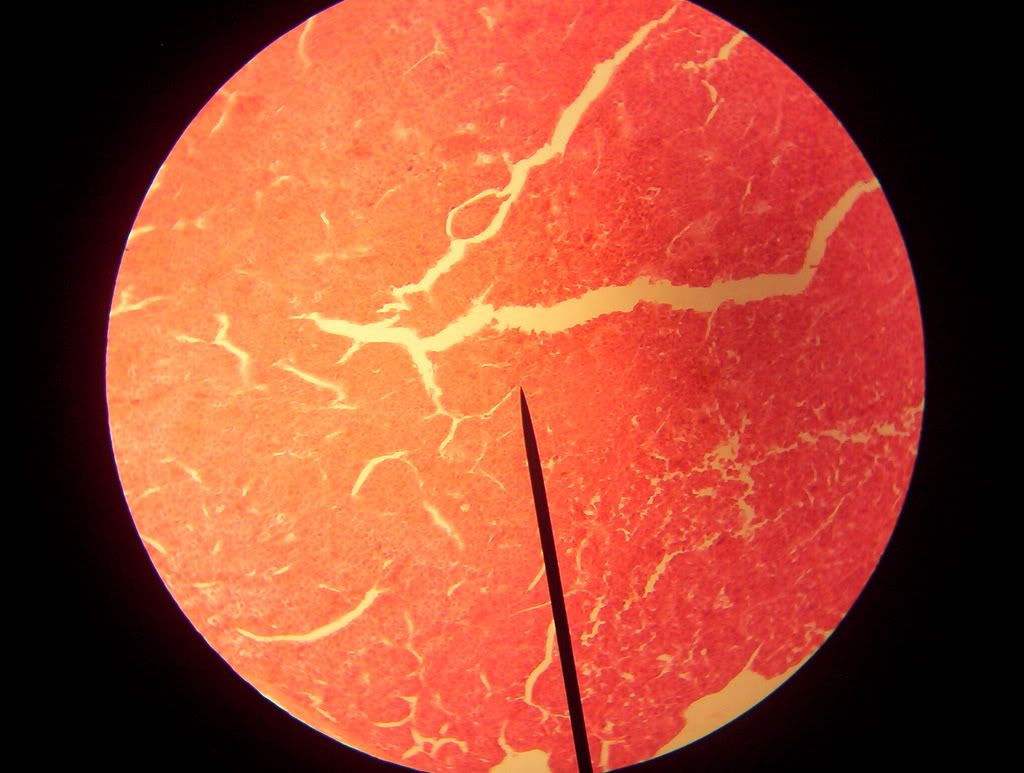 ID: 757630 · |
|
Mr. Majestic Send message Joined: 26 Nov 07 Posts: 4752 Credit: 258,845 RAC: 0
|
Here is what a normal adult lung should look like:  Here is what tuberculosis of the lung looks like:  ID: 757635 · |
AndyW  Send message Joined: 23 Oct 02 Posts: 5862 Credit: 10,957,677 RAC: 18 
|
Albert, love the thread but am concerned that you might have a body hidden in your bedroom that you are hacking up?  ID: 757681 · |
|
Johnney Guinness Send message Joined: 11 Sep 06 Posts: 3093 Credit: 2,652,287 RAC: 0 
|
Albert, Very cool stuff. I'm very impressed! You should mark the slides that you make yourself with something like [MySlide] to distinguish which slides you cut and made yourself. And which ones you bought or were made somewhere else. You obviously are not cutting our your own tonsils to show them to us... LOL. But what about the house fly leg or the human blood cell, is the blood yours?. You can cut slides from plants and insects, but I'm sure it takes time. There is also a tonne of bacteria growing all over food in your fridge, even if you clean it! And yogurt and fresh meat from the butcher! Albert you live in Ohio, so there must be some super cool bugs living in your garden. This is very cool Albert! Very exciting possibilities! What could we splice open and have a look at! John. {EDIT} Tell you what, i don't have a microscope but i have a digi camera. I will take some shots of the bugs here in Ireland and post them. I will see how close i can get with fine detail! {EDIT} I have a massive telescope, but i'm not sure it would do small objects...LOL. Might try the eye peices, they might work! 
ID: 757698 · |
|
Johnney Guinness Send message Joined: 11 Sep 06 Posts: 3093 Credit: 2,652,287 RAC: 0
|
OK, this worked out far better than i thought!. I dismantled the eye pieces of the telescope and taped them onto the front of the digital camera!. But it worked very well! This is the set up i used;  And the samples.....Unfortunately the spider fell to pieces before i took this picture! (Legs detached from the body!)  This sample was a spider that was dead and crusty. Might have died several weeks or months ago! Just take a look at those Fangs!!!   Link for larger image No.3; Image 3 Link for larger image No.5; Image 5 A close up of his eyes!  Link for larger image No.11; Image 11 And here are his legs;   Link for larger image No.7; Image 7 Link for larger image No.8; Image 8 But only when i got close with more powerful lenses, did i see something strange!!!. There was a large hole in his Head!!. This hole was not really visible with the naked eye!     Link for larger image No.10; Image 10 Link for larger image No.15; Image 15 Link for larger image No.16; Image 16 Link for larger image No.17; Image 17 So from forensically examining this spider, i was able to establish that he was actually killed by another spider who left a gapeing hole in this guys head when he bit in with his fangs! I have some more different sample insects that i will do later if i have time! Isn't nature incredible when you look closely:). Remember i done this with telescope lenses taped to the camera ....LOL John.
ID: 757802 · |


©2024 University of California
SETI@home and Astropulse are funded by grants from the National Science Foundation, NASA, and donations from SETI@home volunteers. AstroPulse is funded in part by the NSF through grant AST-0307956.