Mr. Majestic's Microscopy..... and life on Mars?
Message boards :
Cafe SETI :
Mr. Majestic's Microscopy..... and life on Mars?
Message board moderation
Previous · 1 . . . 3 · 4 · 5 · 6 · 7 · 8 · Next
| Author | Message |
|---|---|
 Johnney Guinness Johnney Guinness Send message Joined: 11 Sep 06 Posts: 3093 Credit: 2,652,287 RAC: 0 
|
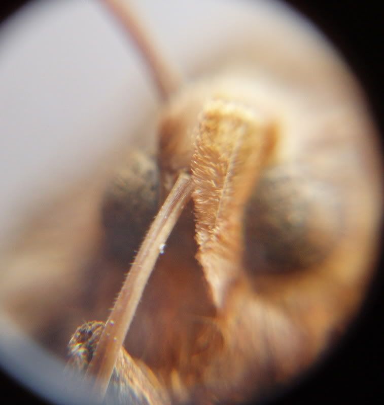
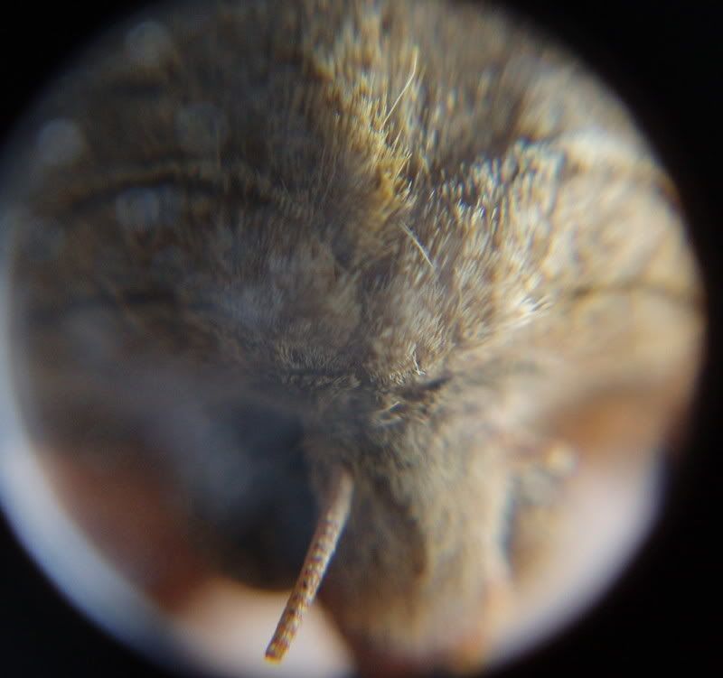
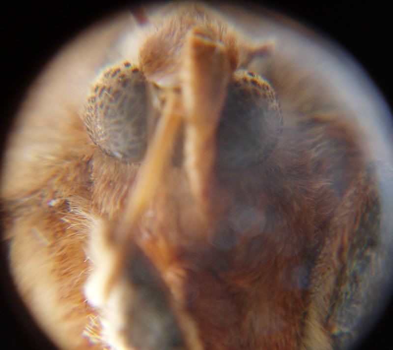
This is a Moth that appeared to have died from exhaustion on my window on a sunny day. Estimated time of death; Several months. The starting sample 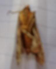 Link for larger image No.1; Image 1 These were the best shots i could get. 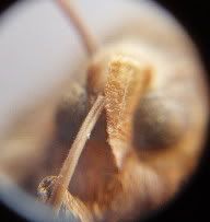 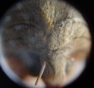  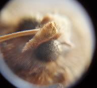 Link for larger image No.2; Image 2 Link for larger image No.3; Image 3 Link for larger image No.6; Image 6 Link for larger image No.10; Image 10 Not as good as the spider shots i took. But not too bad! John. 
ID: 763063 · |
|
Mr. Majestic Send message Joined: 26 Nov 07 Posts: 4752 Credit: 258,845 RAC: 0 
|
I am running out of specimens! If you have any suggestions for things that you would like to see that would be easily obtainable please post them. I plan to order a few more slides later this week, but until them I am low on ideas. Oh, that is a pointer. It is there so if I wanted to point something out in a picture I could rotate the eyepiece so the tip of that would point to what ever it was that I was putting emphasis on. As far as crystals go that's a good idea. @Richard Another good idea. I just have to wait until I can get a hold of one. @Dominique I'm surprised that I didn't think of that one :) Thank you all for your suggestions. What I like about all of these suggestions is that all of them are easily obtainable. I will try to do all of these, though I will probably only have time for the crystals this weekend as I am watching these two all weekend:    ID: 763243 · |
|
Mr. Majestic Send message Joined: 26 Nov 07 Posts: 4752 Credit: 258,845 RAC: 0
|
This is a Moth that appeared to have died from exhaustion on my window on a sunny day. Estimated time of death; Several months. Very interesting post John! I especially like this one: If you don't mind me asking what kind of camera do you use to get these shots? ID: 763244 · |
|
Johnney Guinness Send message Joined: 11 Sep 06 Posts: 3093 Credit: 2,652,287 RAC: 0
|
If you don't mind me asking what kind of camera do you use to get these shots? Its an Olympus u-Mini 5.0 mega pixel. Its a pretty standard camera you would buy for taking family and friends type photos, nothing special. But like i was saying, i use electrical tape to attach parts of the lenses from my telescope to the front of the camera and it works very well. And i have a mini tripod that was part of a spirit level and i have it screwed into the base of the camera to keep it steady.  This is the spirit level, its a laser level! Its rough and ready but it works....LOL Hope them kids don't tare the place up on you while your minding them! John.
ID: 763375 · |
|
Mr. Majestic Send message Joined: 26 Nov 07 Posts: 4752 Credit: 258,845 RAC: 0
|
I never thought of using telescope lenses on a camera, good idea! Now I just have to survive three days of twins wreaking havoc on the house and I will finally have time to experiment with some picture taking. ID: 763419 · |
|
Clyde C. Phillips, III Send message Joined: 2 Aug 00 Posts: 1851 Credit: 5,955,047 RAC: 0
|
I am running out of specimens! If you have any suggestions for things that you would like to see that would be easily obtainable please post them. I plan to order a few more slides later this week, but until them I am low on ideas. All the ideas just come and go from my mind. How about film emulsion? Colloidal dispersions like ink, milk, etc? Computer processor taken apart? CRT, LCD, LED pixels? Hairs, dust, cloth fibers, spider webs? Digital camera sensor? New and used phonograph records and needles? Plastic frosted by oxidation? CD, DVD, Blu-ray disc pits? Video of the movement of the hourhand of a watch? Edge of a new razorblade from different angles? On and on..... ID: 763683 · |
|
Mr. Majestic Send message Joined: 26 Nov 07 Posts: 4752 Credit: 258,845 RAC: 0
|
I am running out of specimens! If you have any suggestions for things that you would like to see that would be easily obtainable please post them. I plan to order a few more slides later this week, but until them I am low on ideas. hmm... a lot of low magnification stuff, I like it! At the moment I am busy with the babies here so it will have to wait until late tonight, but I should have time to do some of these tonight. I am sorry, but the things like the crystals, butterfly wing, pollen, etc. will have to wait until after Sunday when the babies go home. They just seem to like to make a ruckus and it is keeping me quite busy. In fact I better get back to them now.... See you all later! ID: 763776 · |
|
John McCallum Send message Joined: 5 Dec 04 Posts: 877 Credit: 599,458 RAC: 8 
|
 Mouse cell nucleus Mouse cell nucleushttp://news.bbc.co.uk/1/shared/spl/hi/pop_ups/08/technology_enl_1212750145/html/1.stm A detailed 3D image of the nucleus of a mouse cell. The picture was captured using a new imaging microscope technique called three-dimensional structured illumination, described in the journal Science. It allows cell biologists to take high resolution, multi-coloured images. Old enough to know better(but)still young enough not to care ID: 764135 · |
|
Dr. C.E.T.I. Send message Joined: 29 Feb 00 Posts: 16019 Credit: 794,685 RAC: 0
|
. . . notes: regarding the Architecture of a Butterfly Wing Butterfly wing colors are produced by a combination of pigments and reflection from photonic structures. “If you examine the wing scale, you see all of the intricate micron-scale and nanometer-scale features that determine the optical properties,†Wang noted. “From a physical point of view, this is a very regular photonic structure with regular gaps that produce the bluish color.†. . . Replication of Butterfly Wing Scales Provides New Technique for Producing Complex Photonic Structures The artificial butterfly wing scale is a three-dimensional structure that retains the features of the original. That includes hollow tubular structures that split off at regular intervals, providing the potential for use as optical waveguides and optical splitters – and even as microfluidic or microreactor devices . . . This scanning electron microscope image shows the structure on the surface of a butterfly wing scale 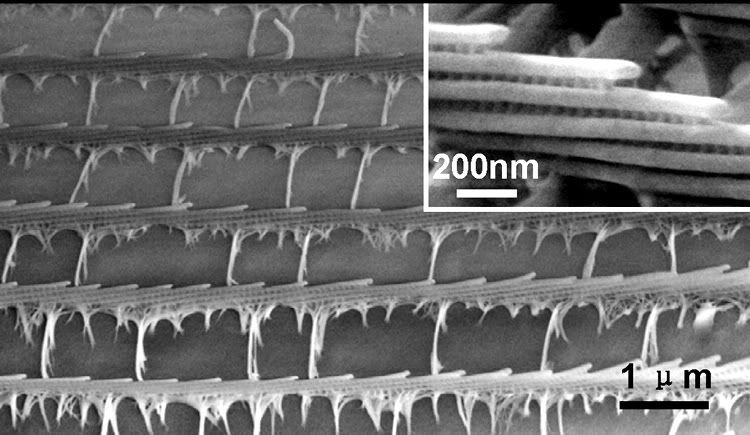 Image courtesy Zhong Lin Wang Optical microscope images of coated butterfly wing scales show color differences related to the thickness of the deposited alumina 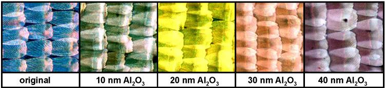 Image courtesy Zhong Lin Wang
Copyright © 2008 - Informations & Photographic Images provided through the courtesy of Georgia Institute of Technology  BOINC Wiki . . . BOINC Wiki . . .Science Status Page . . . ID: 764168 · |
Luke  Send message Joined: 31 Dec 06 Posts: 2546 Credit: 817,560 RAC: 0 
|
I'm not sure how you'd do this, Albert, but could I suggest a "small leaf"? - Luke. ID: 764360 · |
|
Dr. C.E.T.I. Send message Joined: 29 Feb 00 Posts: 16019 Credit: 794,685 RAC: 0
|
I'm not sure how you'd do this, Albert, but could I suggest a "small leaf"? . . . like this?  Figure 2. Bacterial pustule of soybean with (A) upper leaf symptoms, (B) underside of same leaf, and (C) raised pustules on leaf underside (10X magnification). Note lesion association with main leaf veins. ~ or ~ 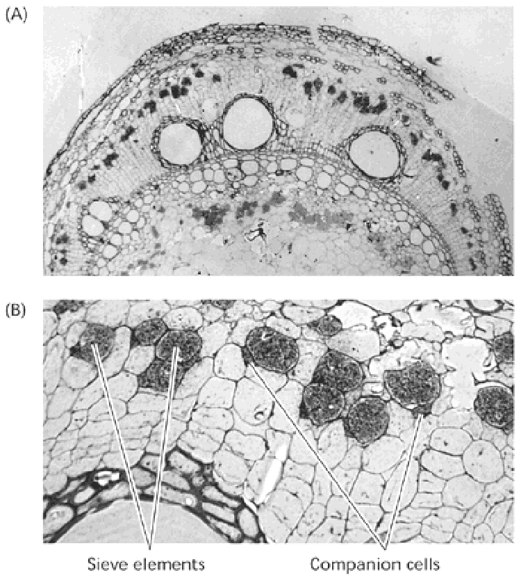 Web Figure 10.1.B Photosynthate from the leaves appears in the sieve elements of phloem in the stem. 14CO2 was supplied to a source leaf of morning glory (Ipomea nil). 14C was incorporated into sugars synthesized in the photosynthetic process, which were then transported to other parts of the plant. The location of the label is revealed in the tissue cross sections by the presence of dark grains on the film. (A) shows a low magnification of the cross section of the stem (50×), revealing dark spots resulting from the silver grains in the film, shown in higher magnification (325×) in (B). The label is confined almost entirely to the sieve elements of the phloem. (Courtesy of D. Fisher.) BOINC Wiki . . .Science Status Page . . . ID: 764400 · |
|
Clyde C. Phillips, III Send message Joined: 2 Aug 00 Posts: 1851 Credit: 5,955,047 RAC: 0
|
It's Over, on your butterfly wings, those electron micrographs look really interesting. However, I have a feeling the the ribs shown on the optical microscope pictures are not the same ribs shown on the electron photos. I've seen butterfly scales with the naked eye and believe they're something like 1/5 mm long (200 microns). Looking at the optical microscope pictures their ribs appear to be about 1/20 the length of the scale or perhaps 10 microns apart. On the electron photos the ribs appear to be only about one micron apart. ID: 764878 · |
|
Mr. Majestic Send message Joined: 26 Nov 07 Posts: 4752 Credit: 258,845 RAC: 0
|
I am sorry to you all to keep you waiting. The babies have just kept me so busy that I haven't had time to keep up with this thread and get some new pics. I hope to have some tonight after they go home, but if not I will get them tomorrow I swear. Thank you to all who has been contributing to this thread and keeping it going, I greatly appreciate it! Take care everyone, John ID: 764921 · |
|
Mr. Majestic Send message Joined: 26 Nov 07 Posts: 4752 Credit: 258,845 RAC: 0
|
On the back of the five dollar bill (America) you will find 26 states listed at the top of the Lincoln memorial: * Arkansas * Michigan * Florida * Texas * Iowa * Wisconsin * California * Minnesota * Oregon * Kansas * West Virginia * Nevada * Nebraska * Colorado * North Dakota * Delaware * Pennsylvania * New Jersey * Georgia * Connecticut * Massachusetts * Maryland * Carolina * Hampshire * Virginia * New York Here is an example of what this looks like: 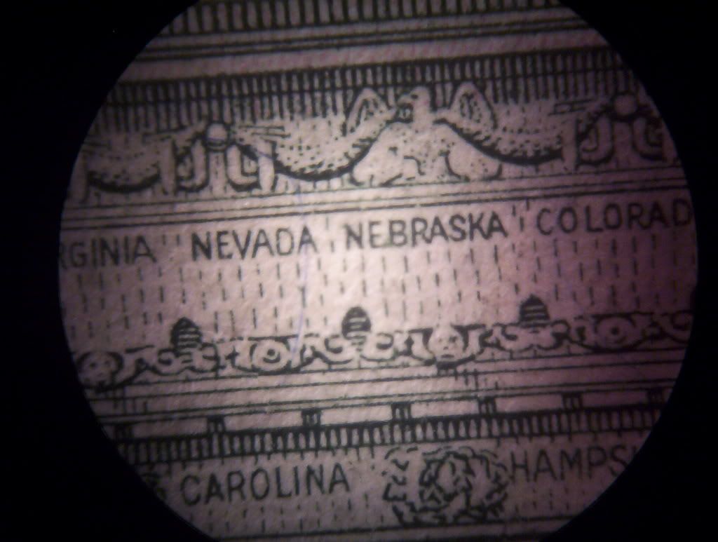 Say hello to Mr.Lincoln: 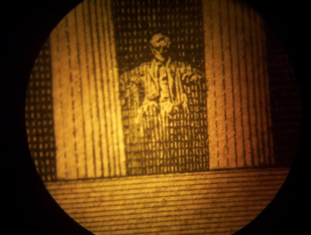 ID: 764960 · |
|
Dr. C.E.T.I. Send message Joined: 29 Feb 00 Posts: 16019 Credit: 794,685 RAC: 0
|
It's Over, on your butterfly wings, those electron micrographs look really interesting. However, I have a feeling the the ribs shown on the optical microscope pictures are not the same ribs shown on the electron photos. I've seen butterfly scales with the naked eye and believe they're something like 1/5 mm long (200 microns). Looking at the optical microscope pictures their ribs appear to be about 1/20 the length of the scale or perhaps 10 microns apart. On the electron photos the ribs appear to be only about one micron apart. . . . eh Clyde - Thank You and here's hopin' You are Well a Note: click on the following Link The cuticular structure in insects is generally investigated by means of SEM and TEM. SEM micrographs of cut surfaces give an impression of the three-dimensional character of the structure, while TEM micrographs of thin sections provide a two-dimensional view > i believe these Factors govern the Observational Differences Noted by you Clyde . . . although, i might even be wrong ;) BOINC Wiki . . .Science Status Page . . . ID: 764997 · |
|
Clyde C. Phillips, III Send message Joined: 2 Aug 00 Posts: 1851 Credit: 5,955,047 RAC: 0
|
I just pulled out a five, found a double-convex lens of about 40mm focal length, found a ruler graduated in 64ths of an inch (0.4mm) and it looked to me that the printing of the states is a little larger than 1/64 inch, perhaps 0.5 millimeter, high. Someday I'll try to catch a butterfly, knock off a few of its scales on a glass and try to measure them in the same way. Celestron finally answered me and said that the 10-power eyepieces of its 44110 have an eye relief of 23.5 millimeters and that its Koehler illuminator has two diaphragms. That's great if it's telling the truth. Maybe I'll order one in a month or so. Maybe someday I'll get a digital camera. That 18x Nikon looks good but I have to be assured that its lens is good, too. Otherwise I'll wait. Terribly-small format lenses might suffer from diffraction, too. ID: 765378 · |
|
Dr. C.E.T.I. Send message Joined: 29 Feb 00 Posts: 16019 Credit: 794,685 RAC: 0
|
I just pulled out a five, found a double-convex lens of about 40mm focal length, found a ruler graduated in 64ths of an inch (0.4mm) and it looked to me that the printing of the states is a little larger than 1/64 inch, perhaps 0.5 millimeter, high. . . . Dear Clyde - i will 'personally' clip YOUR wings - IF - you so much as touch ANY Butterfly scales < jest kiddin' . . . although, i wouldn't 'touch' the Butterfly IF i were you - THEY are quite 'precious' and iT will destroy the 'architecture' - of said wings . . . ;) < 'ello Albert . . . BOINC Wiki . . .Science Status Page . . . ID: 765382 · |
|
Mr. Majestic Send message Joined: 26 Nov 07 Posts: 4752 Credit: 258,845 RAC: 0
|
Hello Richard and clyde. I am sorry for the lack of updates. I have not had the time for them. Tonight I will do them, I swear :) I meant to get a few new samples today on my hike, but I forgot to bring the vials to store them :( If it doesn't rain tomorrow I will go on another hike to get some samples. As for tonight I will try to do some of the requests. Take care, John ID: 765444 · |
|
Mr. Majestic Send message Joined: 26 Nov 07 Posts: 4752 Credit: 258,845 RAC: 0
|
I'm not sure how you'd do this, Albert, but could I suggest a "small leaf"? Here it is at low power: 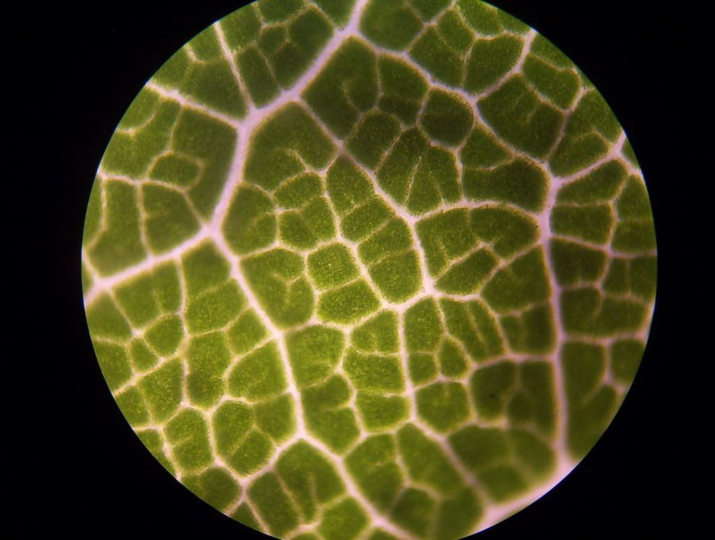 The higher powered view of the cells did not turn out to well because I don't have a microtome to make the proper slides. I had to make the slices with a scalpel, so it is a little thick. For this reason I have the original image and then an image in which I indicated the nuclei with red and the cells with blue Original: 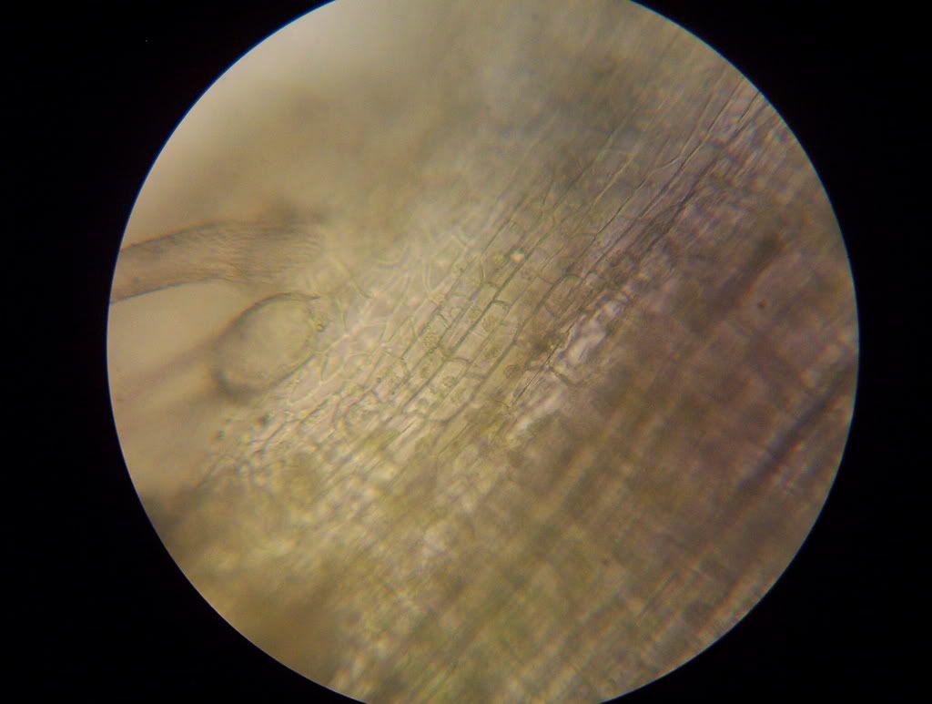 edited: 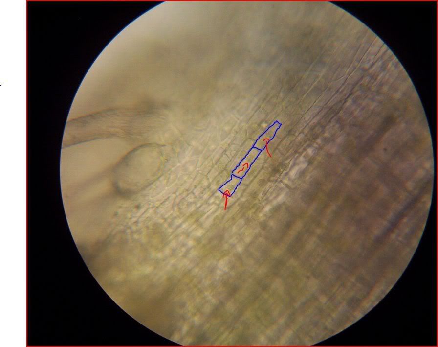 ID: 765551 · |
|
Mr. Majestic Send message Joined: 26 Nov 07 Posts: 4752 Credit: 258,845 RAC: 0
|
Here is the basic structure of a cicada wing, sorry Richard I have not yet gotten a hold of a butterfly wing yet. 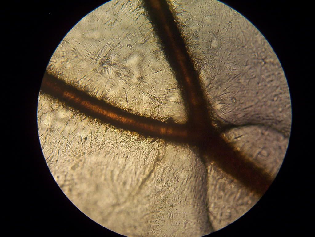 ID: 765552 · |

©2024 University of California
SETI@home and Astropulse are funded by grants from the National Science Foundation, NASA, and donations from SETI@home volunteers. AstroPulse is funded in part by the NSF through grant AST-0307956.